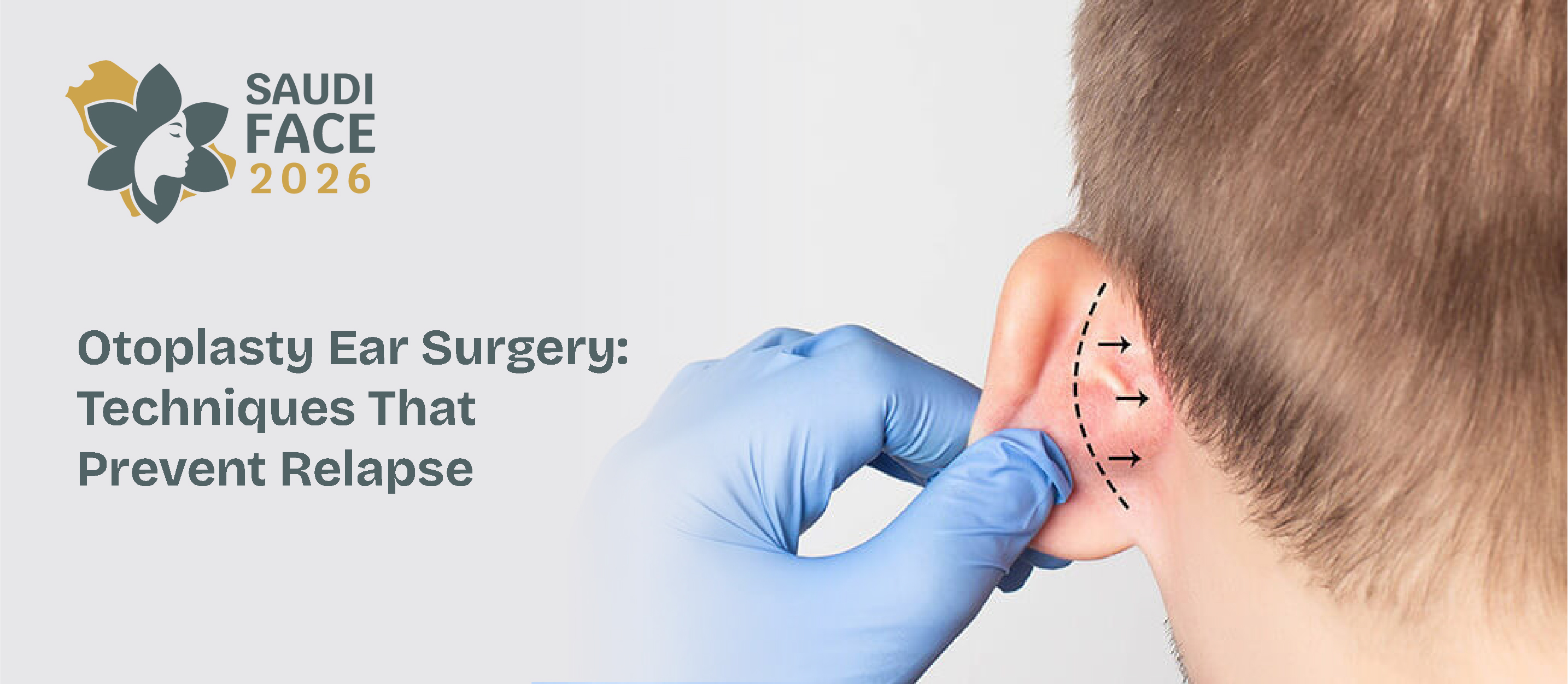
The human ear requires a delicate balance of symmetry, proportion, and contour. Otoplasty addresses congenital or acquired deformities of the external ear, providing profound psychological and aesthetic benefits. This article explores the precise clinical methodologies, surgical techniques, patient safety protocols, and evidence-based recovery strategies essential for modern surgeons performing otoplasty.
What Is Otoplasty?
Otoplasty is a specialized cosmetic and reconstructive surgical procedure designed to alter the size, shape, position, or proportion of the external ear structure. Most commonly utilized to correct prominent ears—a condition frequently stemming from an underdeveloped antihelical fold, conchal hypertrophy, or an increased concho-mastoid angle—the otoplasty procedure combines rigorous anatomical principles with artistic precision.
While the surgical route remains the gold standard for permanent structural remodeling, early infant interventions may leverage non-surgical otoplasty via mechanical molding systems. However, once the ear cartilage calcifies, surgical modification becomes necessary. Financial considerations vary widely; the total otoplasty cost typically reflects the surgeon's expertise, the complexity of the deformity, geographic location, and facility fees. Ultimately, the objective is to establish a natural, harmonious relationship between the ears and the facial profile.
Learn more about Saudi Face 2026
Surgical Techniques for Otoplasty Ear Surgery
Achieving a natural-looking result in otoplasty ear surgery relies on selecting the appropriate technique tailored to the patient's specific structural anomaly. Contemporary otoplasty relies heavily on combinations of cartilage-sparing and cartilage-cutting techniques.
Cartilage-Sparing Techniques (Suture-Based)
-
Mustardé Technique: This foundational technique utilizes permanent, non-absorbable mattress sutures (typically 4-0 clear nylon or polypropylene) placed through the posterior perichondrium and cartilage. This creates or accentuates a deficient antihelical fold.
-
Furnas Technique: To address conchal hypertrophy or an exaggerated concho-mastoid angle, Furnas permanent concho-mastoid sutures anchor the conchal cartilage back to the mastoid periosteum, effectively retropositioning the ear.
Cartilage-Cutting and Scoring Techniques
-
Chongchet and Stenström Techniques: These approaches leverage the Gibson principle—which states that scoring one side of cartilage causes it to warp in the opposite direction. Incising or scoring the anterior perichondrium allows the cartilage to fold backward smoothly, creating a natural antihelical curvature without sharp, artificial edges.
Learn more: Saudi Face: The Leading Plastic Surgery Conference for Facial Aesthetics
Otoplasty Complications - Recognition, Prevention, and Revision Management
Like any delicate aesthetic procedure, otoplasty ear surgery carries inherent risks. Success requires meticulous intraoperative handling and early recognition of postoperative complications.
Early Complications
-
Hematoma: The most critical early complication, marked by severe, disproportionate unilateral pain. Immediate surgical exploration, evacuation, and meticulous hemostasis are mandatory to prevent ischemic cartilage necrosis or infection.
-
Infection and Chondritis: Pseudomonas aeruginosa and Staphylococcus aureus are common culprits. Prophylactic antibiotics are vital. If suppressive chondritis develops, aggressive intravenous antibiotics and potentially surgical debridement are required to protect structural integrity.
Late Complications and Revisions
-
Suture Extrusion: Permanent sutures can spit or erode through thin posterior skin. Treatment involves localized removal of the offending suture, which can usually be done safely if the cartilage has already fibrosed into its new position.
-
Aesthetic Deformities: Overcorrection can lead to a "pinned-back" appearance, or a "telephone ear" deformity where the mid-portion of the ear is over-recessed relative to the helix and lobule. Revision management typically demands scar release, suture adjustment, or cartilage grafting.
Explore the full Saudi Face 2026 agenda and workshop schedule
Pediatric Otoplasty - Surgical Timing, Technique Modifications, and Developmental Considerations
Timing is paramount in pediatric otoplasty ear surgery. The human ear reaches approximately 85% of its adult size by age four to five, and the cartilage transitions from highly malleable to more resilient and rigid.
[Infancy: 0-6 Months] ──> Non-Surgical Otoplasty (Molding) [Preschool: 4-6 Years] ──> Ideal Surgical Window (Cartilage stable, social integration) [Adulthood] ──> Rigid Cartilage (Requires advanced scoring/suturing)
Performing the otoplasty procedure between ages four and six is ideal. At this developmental milestone, the cartilage is sturdy enough to hold sutures securely, yet compliant enough to reshape easily. Furthermore, correcting deformities prior to primary school entry minimizes the profound psychological distress, teasing, and behavioral challenges associated with visible facial differences.
In children, general anesthesia is universally preferred over local sedation. Surgeons must modify their techniques to protect the delicate, thin skin of pediatric patients, ensuring soft-tissue dissection remains atraumatic to preserve optimal blood supply to the growing framework.
Read more: Facial Aesthetics Courses in Riyadh | Injectable Training, Surgical Education
Otoplasty Recovery - Evidence-Based Post-Operative Protocols for Surgeons
An optimized recovery pathway minimizes the overall otoplasty recovery time and protects the newly constructed ear architecture from accidental trauma.
|
Recovery Phase |
Protocol & Patient Instructions |
Clinical Objective |
|
Days 1–3 |
Rigid, non-compressive head dressing; avoid sleeping on sides; strict head elevation. |
Prevent hematoma/seroma formation; minimize edema. |
|
Days 4–14 |
Transition to a soft, supportive headband worn continuously (24/7). |
Maintain ear position; protect against shearing forces. |
|
Weeks 3–6 |
Headband worn exclusively at night; avoid contact sports and strenuous lifting. |
Prevent nocturnal folding or accidental nocturnal avulsion. |
Surgeons should monitor the healing site closely. While initial bruising and swelling resolve within two weeks, the total otoplasty recovery time for complete tissue maturation and scar fading can take up to a full year. Patients should be counseled that mild asymmetry during early healing phases is typical.
Learn more: 5 Skills Every Physician Needs From botox and filler training
Documentation Standards for Otoplasty Before and After
Standardized photographic documentation is a critical medico-legal and clinical requirement for assessing the efficacy of the otoplasty procedure. Capturing high-resolution images allows clinicians to meticulously evaluate structural changes and plan symmetric corrections.
A complete otoplasty before and after photographic series must include the following precise viewpoints:
-
Anterior-Posterior (Full Face): Evaluates overall facial symmetry and the balance of both ears simultaneously.
-
Right and Left Lateral (90-Degree Profiles): Focuses deeply on individual antihelical folds, conchal depth, and helical rims.
-
Oblique Views (Three-Quarter): Showcases the depth and projection of the ears relative to the malar and temporal regions.
-
Close-Up Posterior Views: Documents the postauricular sulcus and provides an essential baseline for tracking incision healing.
Consistency in lighting, background, and patient positioning is non-negotiable to track true structural transformations.
Meet the international facial aesthetics faculty at Saudi Face 2026
Frequently Asked Questions About Otoplasty for Surgeons and Specialists
1. What are the key surgical steps in the otoplasty procedure and how is ear cartilage reshaped?
The otoplasty procedure typically begins with a postauricular incision to access the underlying cartilage matrix. The soft tissue is carefully dissected to expose the perichondrium. Cartilage reshaping is accomplished using a customized combination of Mustardé mattress sutures to reconstruct the antihelical fold and Furnas concho-mastoid sutures to set back an enlarged concha. If the cartilage is rigid, precise anterior scoring is executed to weaken the cartilage's spring, forcing it to bend smoothly without breaking. Excess skin or soft tissue within the postauricular sulcus may be excised before closing the incision with absorbable or non-absorbable sutures.
2. What are the most common complications of otoplasty ear surgery and how are they managed?
The most common complications of otoplasty ear surgery include post-operative hematomas, localized skin or cartilage infections, asymmetric relapse, and suture extrusion. A hematoma demands immediate surgical drainage. Infections require prompt empiric antibiotic therapy targeted at skin flora and Pseudomonas species. Asymmetric recurrence or severe contour deformities (like the telephone ear) must be managed through revision surgery, typically delayed for at least six months to allow local tissues to fully heal and stabilize.
3. Can a non-surgical otoplasty yield permanent results for adults?
No, non-surgical otoplasty using ear molding systems is only effective during the first few weeks to months of life. In infants, high circulating levels of maternal estrogen keep the ear cartilage extraordinarily pliable. As the child grows, the cartilage calcifies and hardens. In adults, non-surgical approaches are ineffective for permanent structural corrections, making traditional surgical intervention the only reliable option.
4. What factors determine the overall otoplasty cost?
The comprehensive otoplasty cost is dictated by several variables: the complexity of the specific ear deformity, the type of anesthesia used (local versus general), operating room or facility fees, and the geographic location of the practice. Surgeons should provide patients with a transparent breakdown of fees during the initial consultation to ensure clear expectations.
.png)
.png)
.png)
.png)
